THE DAILY DAIRY GAME\\ 09-05-2024 // by @rad-austine
Hi friends, greetings to you all, I hope you all are in good health as I am strong and healthy, it's a pleasure to you with you all on my diary game for today Thursday 09-05-2024 in today's activity am going to discuss how I carry out a routine medical chest x-ray examination.
My activity for today was very strenuous I conducted a lot of medical X-ray examinations, but to be precise chest X-rays were more than other types.
X-ray machine
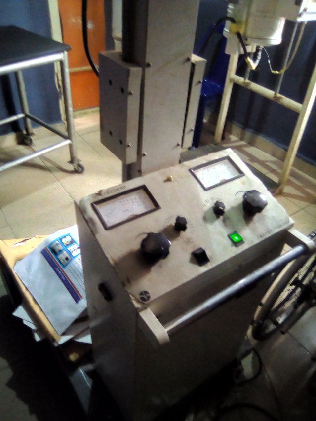
The diagnostic X-ray machine is used to examine the bone in the body to detect if there is a fracture and to examine joint space for osteoporosis, osteoarthritis, dislocation, etc it is also for the examination of internal organs such as lungs heart, etc.
The importance of xx-rays is so numerous to be mentioned because it is the basic modality upon which other modalities are built such as MRI CT scans etc all follow the basic principles of X-ray.
As a Medical Radiographer carry out x-ray examinations every day for various types of patients, such as inward patients outdoor patients (ambulance patients, and emergency patients for accident and surgery cases.
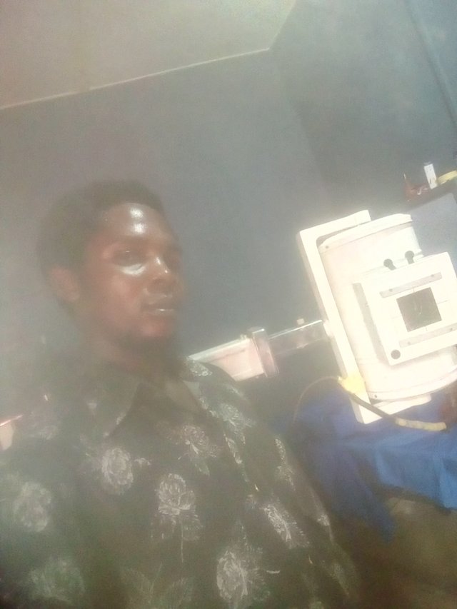
After registering of patients for the type of examination requested I went to my X-ray machine and did what is known as tube warming, which selects my factors depending on the mass of the patient
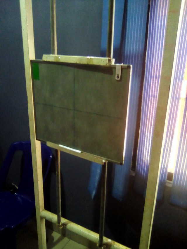
Erect bucky, x-ray cassette, and anatomical marker
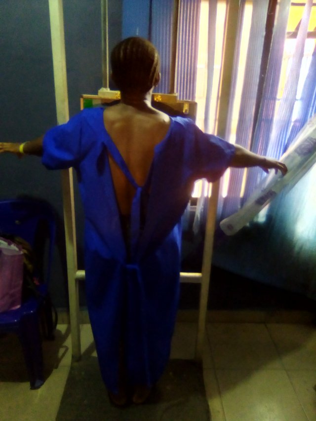
Patient position on x-ray cassette
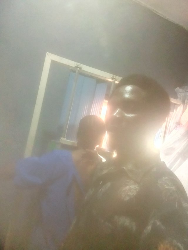
Myself and patient ready for x-ray exposure
I Called the patient to the X-ray room I gave my patient changing instructions to take off her dress and put on the gern the patient stood on the erect bucky for a chest X-ray, I then positioned the patient and gave her breathing instructions which was deep inspiration before exposure after finish positioning the patient
I then move to the X-ray tube head and adjust it to be in correspondence with the radiographic landmark for the chest X-ray is usually T6 after that set the light beam diaphragm collimate it to include the thorax for chest X-ray all the areas of the interest most be capture.
I then move over to the control panel to check my exposure factors such as kv, ma ffd, and sec. I then instructed the patient to breathe into finally exposed the patient.
The effect of radiation dose on the body is that it can cause cancer, so too much radiation dose is not good that is the main reason X-rays are not done on pregnant women, and a good diagnostic X-ray facility should be well sshieldedwith lead to protect against radiation dose.
Finally, after exposure of the patient I then process the image into a radiograph in the darkroom through Manuel processor it's difficult because it Manuel i put the film in to the developer for about 3 to 5 minutes then put it in rinse then fixer for about 5minute finally i wash the film the image is now called a Radiograph then i dry the film for about 30minute after drying.
i then view the Radiograph in a viewing box for interpretation of the Radiograph an the patient was having pneumonia.
I hope you found my diarygame captivating and you learn from it.
10% of this post reward is donated to @hotnewscommunity
Thanks you all for going through my post
Thank you for publishing a post on the Hot News Community, make sure you join at least #club5050, don't plagiarize, use original photos or copyright-free images by linking the source
Moderation note: Keep sharing your best posts and interact with each other in the comments.
Verified by : @fantvwiki