Peripheral Blood Smear (Lab Diaries)
A patient presents to the hospital with complaints of tiredness and fatigue. On examination, she appears to be jaundiced as well as anemic. After a few questions, the doctor writes out a form for lab investigations - a complete blood count and a peripheral blood smear.
Peripheral Blood Smear
We had this as part of our practicals in hematology yesterday. Blood smears are done mainly to identify problems with the blood cells, such as altered shape, size or number.
A blood smear is a drop of blood which is spread on a slide. The blood sample is collected commonly from the arm (peripheral blood vessels).
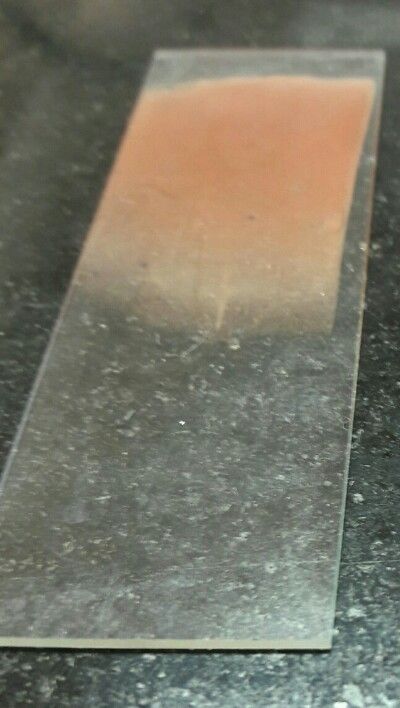
Following preparation of the smear, it is stained. Normal tissues are invisible under a microscope, hence to make them visible, dyes called used, called stains.
We used Leishman stain, which falls under the Romanowsky group of stains. It contains an acidic and basic dye and is used to stain WBCs (white blood cells involved in immune response).
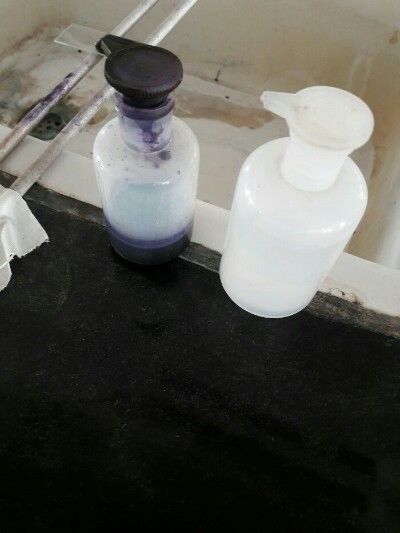
The smear is covered with the stain and left for about 2 minutes during which the cells are 'fixed' onto the slide by methyl alcohol, a component of the stain.
Fun fact - Drinking methanol can kill you. Following ingestion, it is metabolized into formaldehyde and then into formic acid/ formate. Formic acid inhibits cytochrome c oxidase leading to cellular hypoxia and also causes metabolic acidosis. If untreated, it can lead to death.
The stain is then diluted with distilled water (twice the amount of stain used) and left for 15-20 minutes. The stain is washed off and the smear is ready to be observed under microscope. After adding a drop of oil for the oil immersion view.
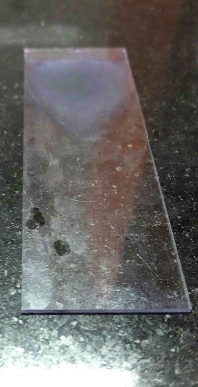
(Sadly, I couldn't get a picture of the microscopic view)
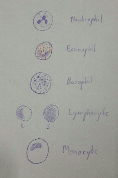
The different white blood cells appear (normally) as follows:-
Neutrophils - Largest in number, lobed nucleus
Lymphocytes - High nucleus to cytoplasm ratio; two types - small and large.
Basophils - Heavily granulated, nucleus not visible.
Eosinophils - Orange granules
Monocytes - Large cells with kidney shaped nucleus, no granules.
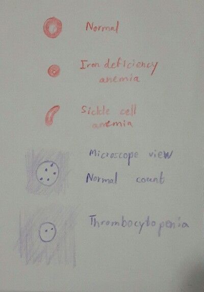
Now onto the practical uses. As I said earlier, its used mainly for identifying blood disorders. The number, size or shape of cells may be changed in certain diseases/conditions, thus helping to identify them.
Some of the identifiable disorders include sickle cell anemia, megaloblastic anemia, thrombocytopenia (decreased platelet count) among others.
Sources:
Lecture notes I took
Pics by me
Sketches by me

Thanks @rocking-dave for the GIF :)