Atomic Force Microscopy of a Gallium Thin Film - Scientific Report
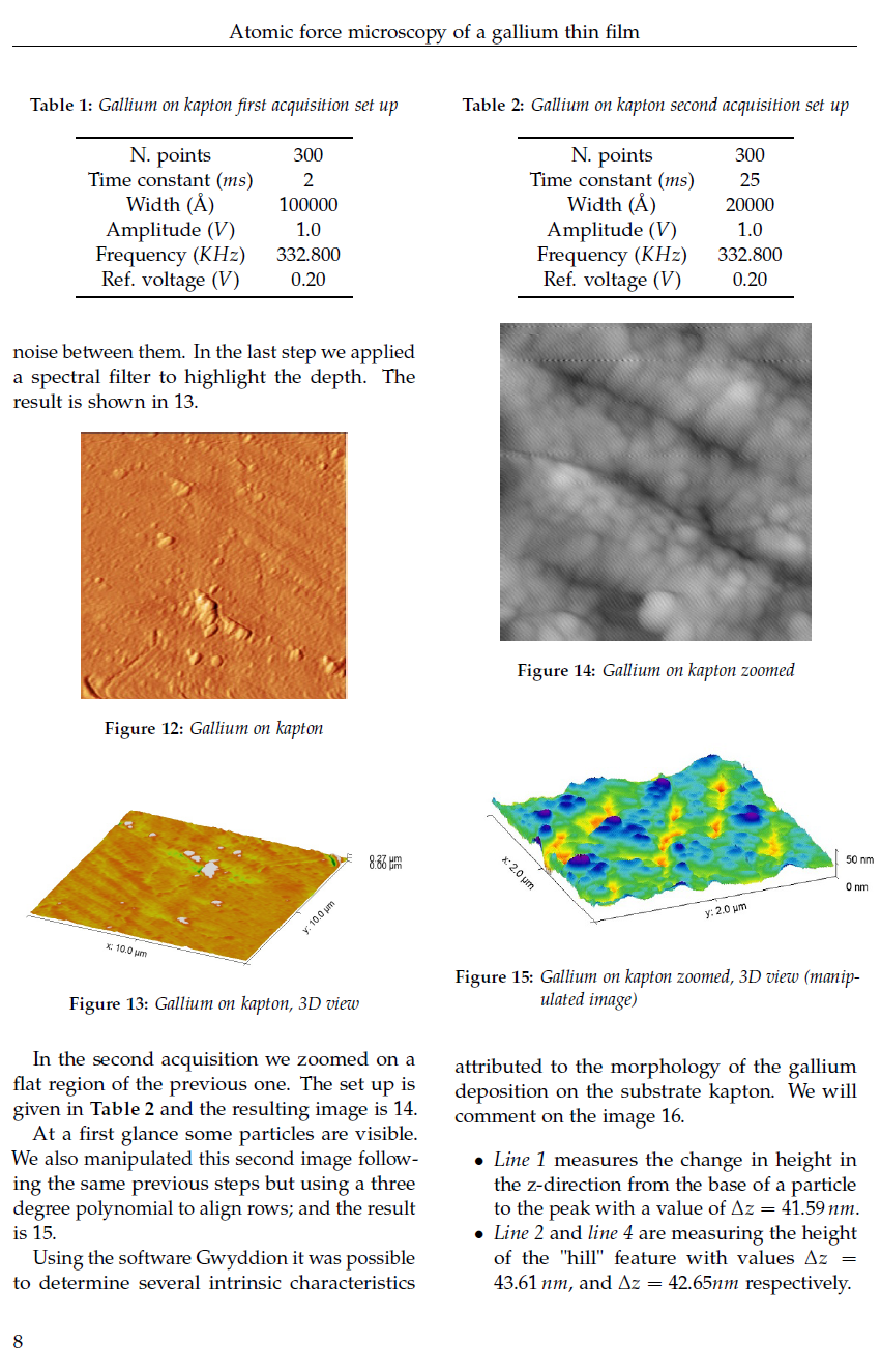
A Scientific Report On Atomic Force Microscopy Performed on Gallium. Learn About AFM And How Gallium Looks At The Atomic Level.
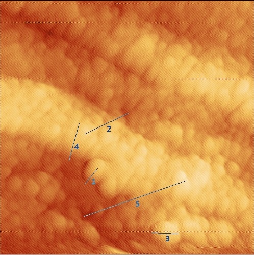
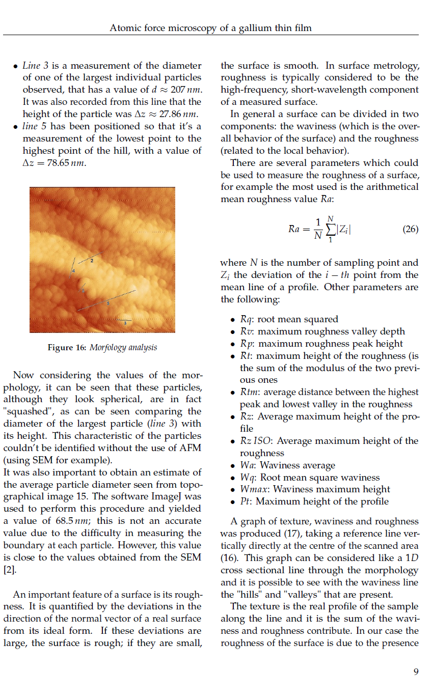
This is an image at the that shows small nano-spheres of Gallium. Line 1 measures the change in height from the base of a particle to the peak with a value of 41.59 nano meters. Line 2 and line 4 are measuring the height of the "hill" feature with values 43.61 nano meters, and 42.65 nano meters respectively. Line 3 is a measurement of the diameter of one of the largest individual particles observed, that has a diameter of 207 nano meters. It was also recorded from this line that the height of the particle was 27.86 nano meters.

Quick Overview
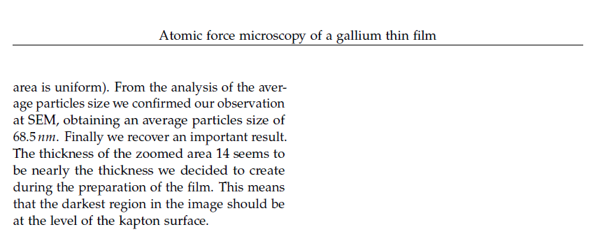
This article is different from my usual style. As steemit doesn't support latex syntax I can't express values as I would like to. So this consists of snap shots of my PDF document. You'' find out exactly what is Atomic Force Microscopy and see some actual results that me and my team obtained in the laboratory. The image you see above is an image produced of the surface of a Gallium oxide. We evapourated Gallium metal and allowed it evapourate on different materials, we did it really slowly so the thickness of the Gallium is at the nano meter scale. Being a science nerd, when I saw this data and produced the image I was "buzzing", You'll understand the sensitivity by reading this article. You'll also have the chance to understand what the results mean. It's quite advanced, but what I'll say to those who are worried about understanding is that it's explained well and clear, don't be conserned about the equations, but you'll understand thier physical meaning. Give it a chance, have a read and see what you learn. If you're not fussed about the physics and maths and just want to see the results, then go to that section and have a look what the atomic scale looks like.
The article follows a page like format, read the left colum then the right column of each individual page. Not that you can't read, just incase you wonder what's going on. :D
Physics.Benjamin
If you liked this post feel free to UPVOTE, FOLLOW, and RESTEEM.

References:-
[1] V.L. Mironov, "Fundamentals of scanning
probe microscopy".
[2]Me and my three friends paper:-"Raman and SEM investigation
of gallium metal"
I will post this paper tomorrow
All images are Creative Commons or public domain, no copyright infringements have occurred. These were contained within a scientific report.
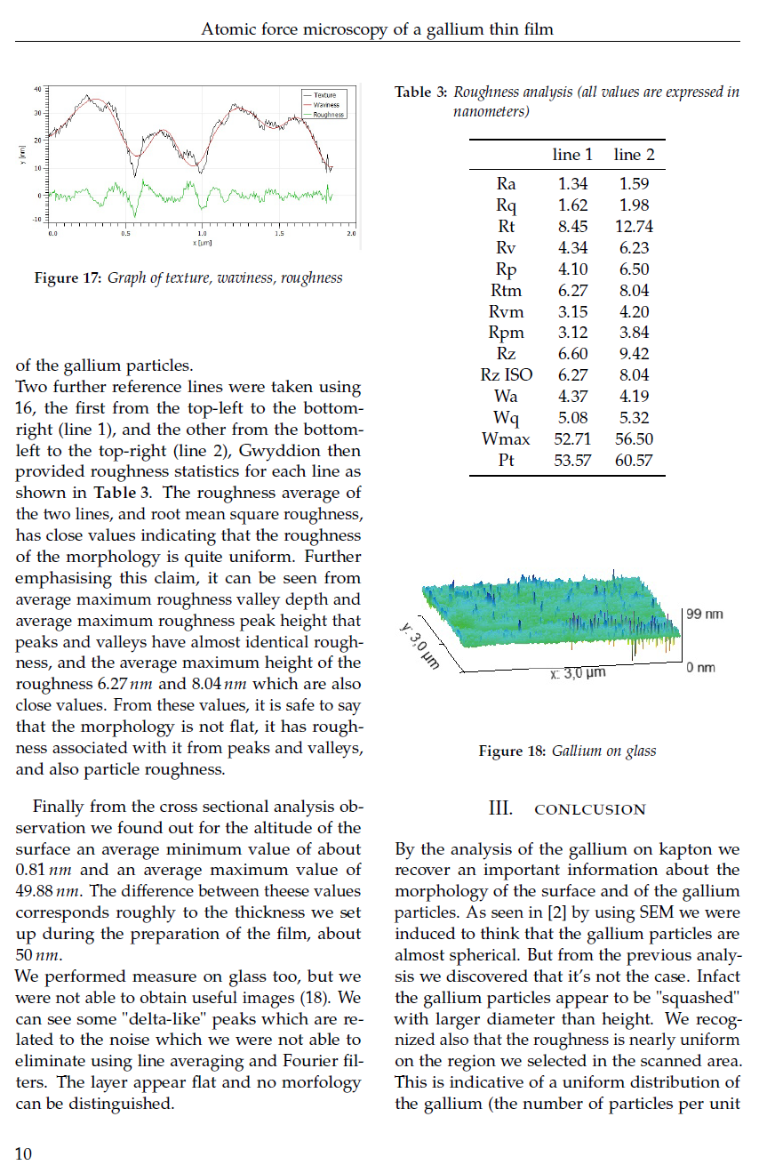
Interesting article @physics.benjamin
The galium surface image is really nice, well I also use AFM but we generally do it to see the peptide nano structure that self assembles and form several structures..
Cheers