Inside the Moonshot Effort to Finally Figure Out the Brain
AI is only loosely modeled on the brain. So what if you wanted to do it right? You’d need to do what has been impossible until now: map what actually happens in neurons and nerve fibers.
"Here’s the problem with artificial intelligence today," says David Cox. Yes, it has gotten astonishingly good, from near-perfect facial recognition to driverless cars and world-champion Go-playing machines. And it’s true that some AI applications don’t even have to be programmed anymore: they’re based on architectures that allow them to learn from experience.
Yet there is still something clumsy and brute-force about it, says Cox, a neuroscientist at Harvard. “To build a dog detector, you need to show the program thousands of things that are dogs and thousands that aren’t dogs,” he says. “My daughter only had to see one dog”—and has happily pointed out puppies ever since. And the knowledge that today’s AI does manage to extract from all that data can be oddly fragile. Add some artful static to an image—noise that a human wouldn’t even notice—and the computer might just mistake a dog for a dumpster. That’s not good if people are using facial recognition for, say, security on smartphones.

A technician observes the brain of a live rat during a test.
To overcome such limitations, Cox and dozens of other neuroscientists and machine-learning experts joined forces last year for the Machine Intelligence from Cortical Networks (MICrONS) initiative: a $100 million effort to reverse-engineer the brain. It will be the neuroscience equivalent of a moonshot, says Jacob Vogelstein, who conceived and launched MICrONS when he was a program officer for the Intelligence Advanced Research Projects Agency, the U.S. intelligence community’s research arm. (He is now at the venture capital firm Camden Partners in Baltimore.) MICrONS researchers are attempting to chart the function and structure of every detail in a small piece of rodent cortex.
It’s a testament to the brain’s complexity that a moonshot is needed to map even this tiny piece of cortex, a cube measuring one millimeter on a side—the size of a coarse grain of sand. But this cube is thousands of times bigger than any chunk of brain anyone has tried to detail. It will contain roughly 100,000 neurons and something like a billion synapses, the junctions that allow nerve impulses to leap from one neuron to the next.
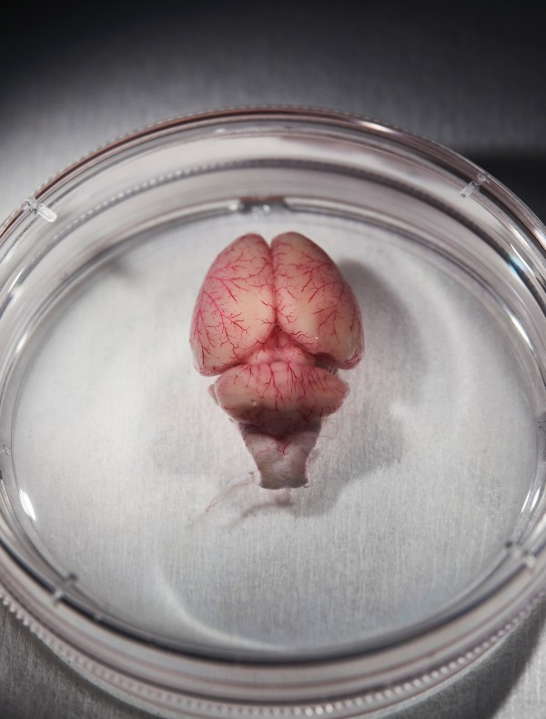
A rat brain in a dish
It’s an ambition that leaves other neuroscientists awestruck. “I think what they are doing is heroic,” says Eve Marder, who has spent her entire career studying much smaller neural circuits at Brandeis University. “It’s among the most exciting things happening in neuroscience,” says Konrad Kording, who does computational modeling of the brain at the University of Pennsylvania.
Zooming in
he MICrONS teams—one led by Cox, one based at Rice University and the Baylor College of Medicine, and a third at Carnegie Mellon—are each pursuing something that is remarkably comprehensive: a reconstruction of all the cells in a cubic millimeter of a rat’s brain, plus a wiring diagram—a “connectome”—showing how every cell is connected to every other cell, and data showing exactly which situations make neurons fire and influence other neurons.
The first step is to look into the rats’ brains and figure out what neurons in that cubic millimeter are actually doing. When the animal is given a specific visual stimulus, such as a line oriented a certain way, which neurons suddenly start firing off impulses, and which neighbors respond?
As recently as a decade ago, capturing that kind of data ranged from difficult to impossible: “The tools just never existed,” Vogelstein says. It’s true that researchers could slide ultrathin wires into the brain and get beautiful recordings of electrical activity in individual neurons. But they couldn’t record from more than a few dozen at a time because the cells are packed so tightly together. Researchers could also map the overall geography of neural activity by putting humans and other animals in MRI machines. But researchers couldn’t monitor individual neurons that way: the spatial resolution was about a millimeter at best.
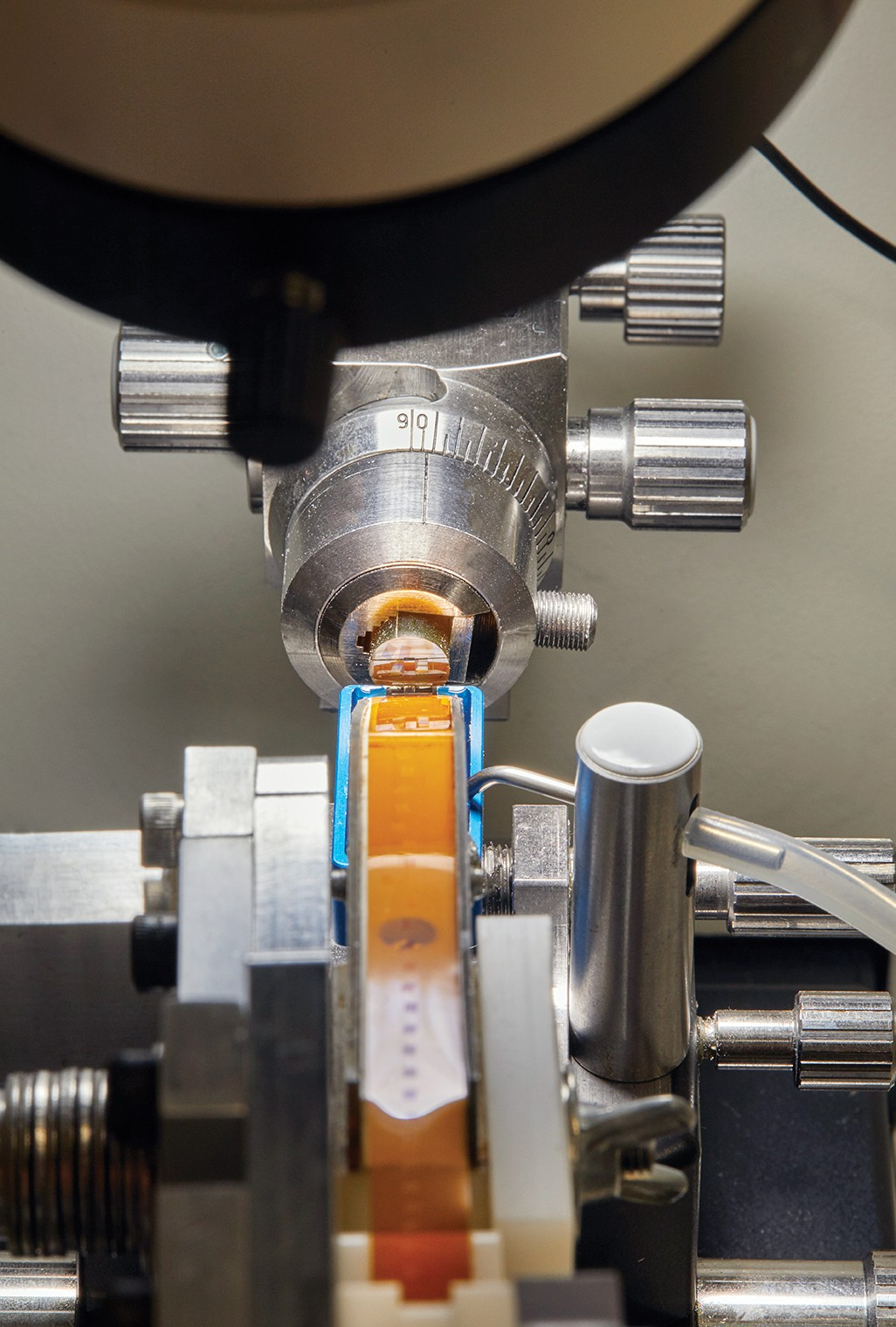
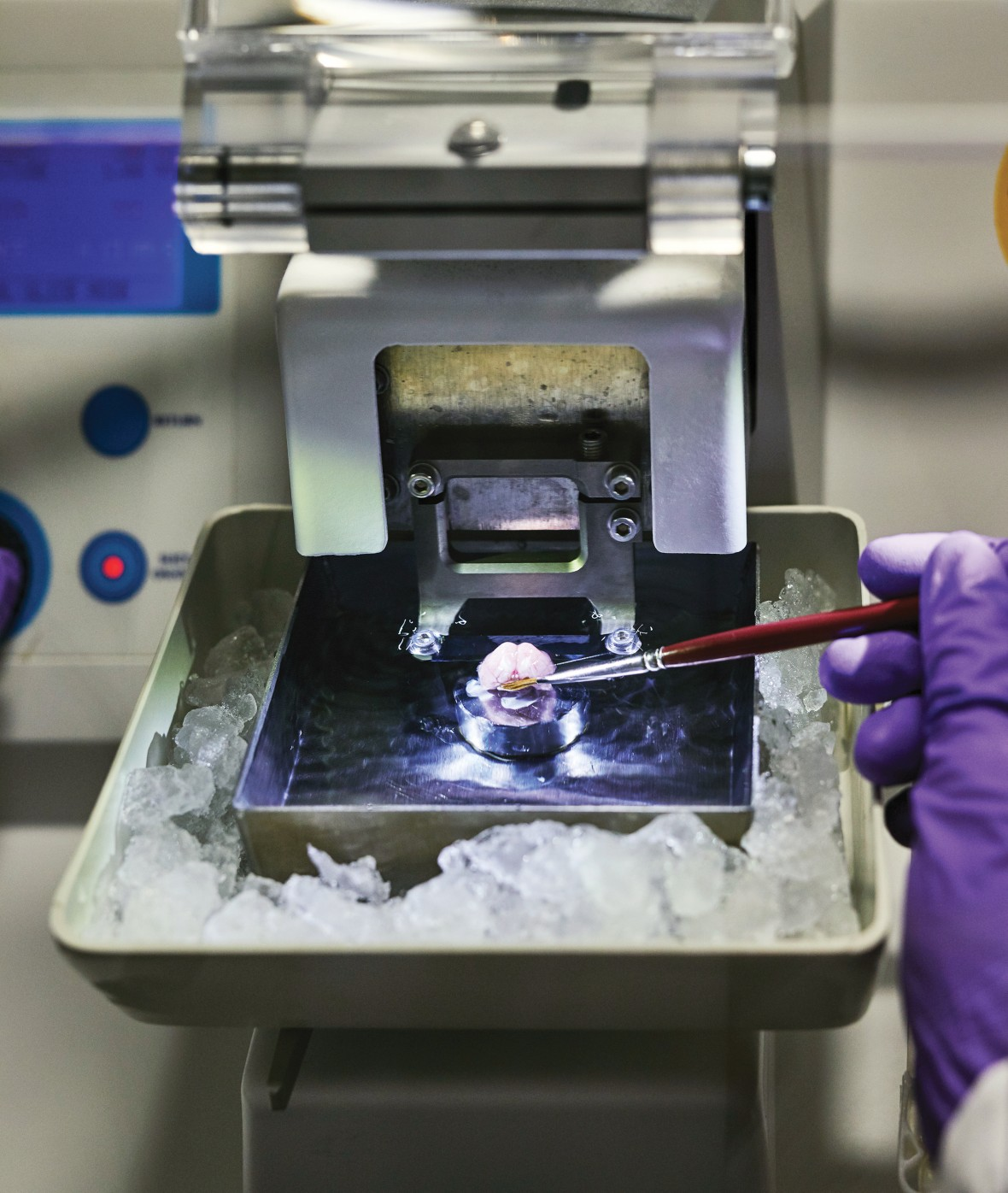
The cut slices of brain stick to a plastic tape.
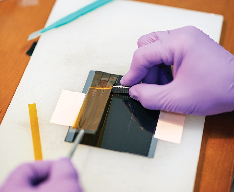
The tape, with brain samples attached, is trimmed and put on a slide plate that will go into a huge scanning machine.
wowwwwwwww
thank you
Hi! I am a robot. I just upvoted you! I found similar content that readers might be interested in:
https://www.technologyreview.com/s/609070/inside-the-moonshot-effort-to-finally-figure-out-the-brain/
I'm so sorry for that
It was nice post just cary on
nice