Understanding the Electrocardiogram (ECG)
An Electrocardiogram (ECG) is a simple, non-invasive diagnostic test used to record the electrical activity of the heart. It plays a vital role in modern healthcare by helping healthcare professionals assess heart rhythm, heart rate, and the overall function of the cardiac conduction system. ECGs are commonly performed in hospitals, clinics, emergency departments, and ambulances to detect heart abnormalities and guide treatment decisions.
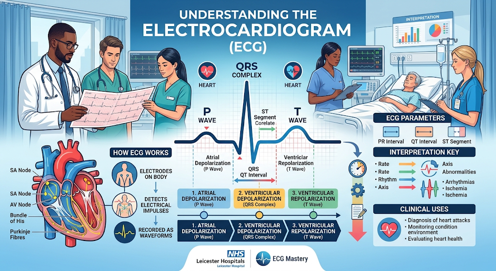
The heart generates electrical impulses that travel through a specialized conduction pathway. The impulse begins at the sinoatrial (SA) node, the heart's natural pacemaker, causing the atria to contract. The signal then passes through the atrioventricular (AV) node, Bundle of His, bundle branches, and Purkinje fibres, leading to ventricular contraction. An ECG captures these electrical events through electrodes placed on the patient's skin.
A standard 12-lead ECG consists of ten electrodes positioned on the limbs and chest. These electrodes provide twelve different views of the heart's electrical activity, allowing clinicians to identify abnormalities from various angles. The main components of an ECG trace include the P wave, which represents atrial depolarisation; the QRS complex, which represents ventricular depolarisation; and the T wave, which represents ventricular repolarisation. The PR interval measures conduction from the atria to the ventricles, while the QT interval reflects the total time required for ventricular depolarisation and repolarisation.
ECGs are invaluable in diagnosing a wide range of cardiovascular conditions. They can identify arrhythmias such as atrial fibrillation, atrial flutter, bradycardia, tachycardia, and heart blocks. ECGs are also essential for detecting myocardial infarction (heart attack), myocardial ischaemia, electrolyte imbalances, and the effects of certain medications. Furthermore, they can help monitor patients with pacemakers and evaluate the effectiveness of treatments.
The procedure is quick, painless, and typically takes only a few minutes. Patients are asked to remain still during recording to reduce interference and ensure an accurate result. Although ECGs provide important information about cardiac electrical activity, findings should always be interpreted alongside the patient's symptoms, medical history, physical examination, and other diagnostic investigations.
In conclusion, the ECG is one of the most important tools in cardiovascular assessment. Its ability to rapidly identify potentially life-threatening cardiac conditions makes it indispensable in both emergency and routine clinical practice, contributing significantly to patient diagnosis, monitoring, and treatment planning.